Case Quiz (October 2023)
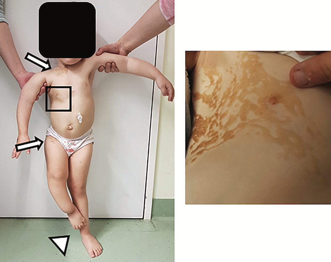
WA female toddler, aged three, was directed to our outpatient nephrology clinic due to a left-sided multicystic dysplastic kidney. Upon physical examination, the patient exhibited several clinical abnormalities, with the most notable being a linear, partially ulcerated nevus sebaceous spanning the entirety of the right upper thoracic cage (Figure).
Moreover, we observed a hemangioma measuring 0.7 x 0.5 x 0.3 cm near the right mandible. Additional characteristics comprised uneven and delayed growth, along with a leg length discrepancy, as well as a thoracic deformity exhibiting scoliosis. The patient's medical history unveiled persistent, widespread bone pain that was severe in intensity, impacting her entire body and severely restricting her mobility,
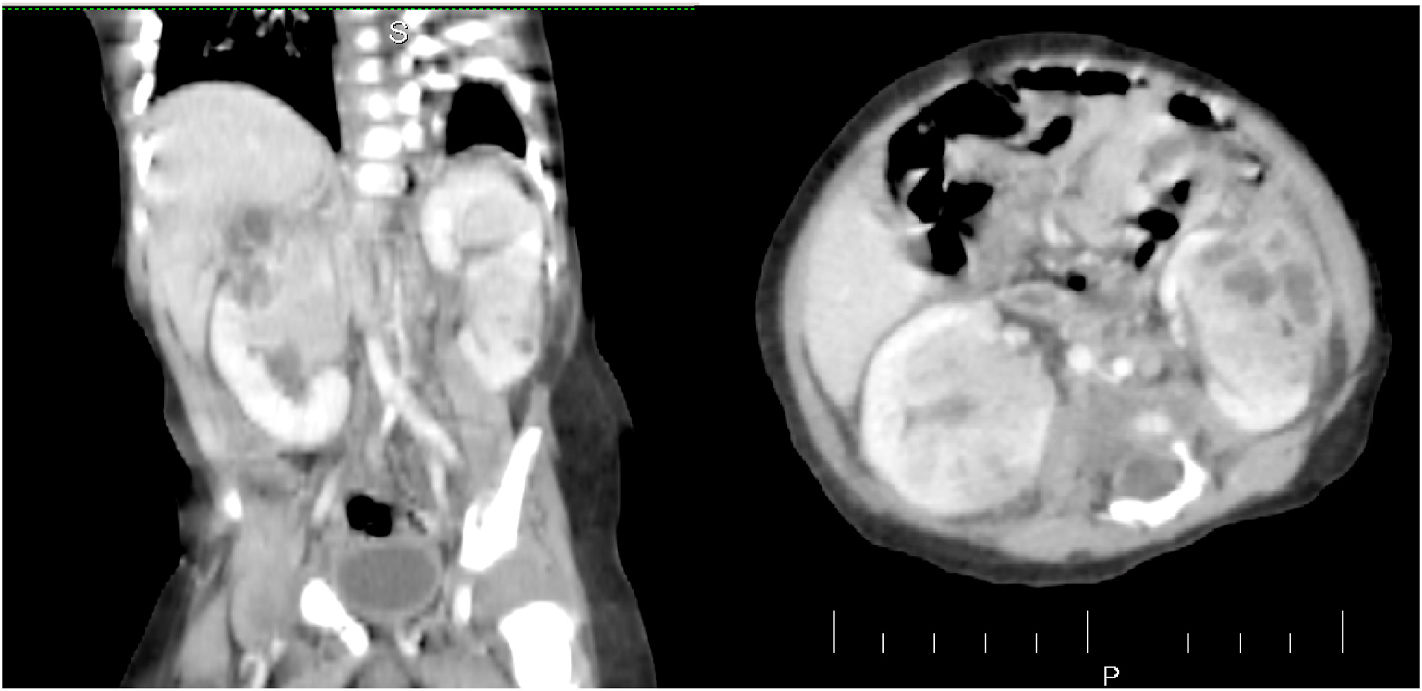
The initial blood analysis indicated significantly reduced phosphate levels 2.1 mg/dl. 25 (OH) D3 levels were notably low at 13 ng/ml, while FGF23 levels were markedly elevated at >35,000 RU/ml (normal range: 25-110 RU/ml). Osteocalcin and calcium fell within the appropriate range for the patient's age. Parathyroid hormone was elevated at 14 pmol/l (normal range: 1.6-6.9 pmol/l). Bone-specific alkaline phosphatase was significantly elevated at 754 mg/l. Renal function parameters, including creatinine, urea, and glomerular filtration rate, were within normal limits. Urine analysis indicated increased phosphate excretion. Radiographic examination showed a delicate, under-mineralized bone structure with a loss of definition at the calcification zone in the epiphyseal-metaphyseal interface and disorganization of the growth plate (Figure). Height measurements consistently fell below the third percentile (height: 78.3 cm, SDS -4.7), while bone age corresponded to that of a one-year-old (at the age of three). Abdominal ultrasound confirmed the presence of a left-sided multicystic dysplastic kidney, without any other intra-abdominal malformations.
Following a bout of Legionella pneumonia, our patient developed a persistent, chronic need for oxygen. A CT scan of the lungs affirmed the severe thoracic deformity and additionally revealed widespread consolidation of the lung parenchyma, accompanied by ground glass opacifications in both lower lobes. To exclude neurological abnormalities, we performed a cranial MRI, revealing a single small arachnoid cyst as an incidental finding without further necessity for intervention.
A diagnostic test was done.


Case Answer (October 2023)
Cutaneous-skeletal hypophosphatemia syndrome (CSHS)
CSHS is characterized by presence of epidermal nevi and elevated levels of FGF-23, leading to hypophosphatemic rickets accompanied by bone pains, fractures, scoliosis and limb deformities.
The combination of clinical and diagnostic findings, except lung involvement, was suggestive for an underlying epidermal nevus syndrome. A biopsy of epidemal nevus was performed for histological and genetic examination.
The unilateral multicystic kidney could be an incidental finding and unrelated to CSHS.
